



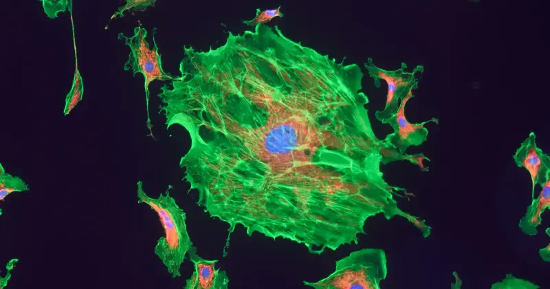
Inside a healthy human muscle fibre, a mitochondrion the length of a bacterium is churning out ATP by pumping protons across a membrane only 7 nanometres thick — and when that membrane starts to leak, cellular sensors flag it within minutes, tag it with a protein called PINK1, and hand it over to a recycling crew that dismantles the entire organelle before it can poison the neighbourhood. The process is called mitophagy, and biologists at institutions from Cambridge to the Pennington Biomedical Research Center have spent the last decade mapping exactly how a cell decides when one of its own power plants has crossed the line from repairable to lethal.
The stakes are not abstract. A single muscle cell can hold more than a thousand mitochondria. A neuron in the substantia nigra, the brain region that fails first in Parkinson’s disease, holds several thousand. If even a fraction of them start leaking and are not cleared in time, the cell around them begins to corrode from the inside.
Why a broken mitochondrion is worse than no mitochondrion at all
A functioning mitochondrion runs an electron transport chain — a series of four protein complexes embedded in its inner membrane that pass electrons down an energy gradient, using the released energy to pump protons and ultimately synthesise ATP. The system is efficient but it is not clean. Even in a healthy organelle, roughly 0.1 to 2 percent of the electrons escape and react with oxygen to form superoxide, the parent molecule of a family called reactive oxygen species.
A damaged mitochondrion loses that discipline. Membrane potential drops. Electrons back up in the chain. The leak rate rises. Hydrogen peroxide, hydroxyl radicals and peroxynitrite pour out into the cytoplasm, oxidising nearby proteins, cutting DNA strands, and cross-linking lipids in the surrounding membranes. Researchers describe the pattern of damage in dry language — controlled utilisation of reactive oxygen species tipping into uncontrolled release — but the mechanical image is closer to an engine slowly clogging on its own exhaust, and then venting that exhaust into the passenger cabin.
The insult compounds. Oxidised lipids in the mitochondrial membrane become more permeable, which drops the potential further, which raises the leak rate again. Within the organelle, a cardiolipin molecule on the inner membrane flips to the outer surface, exposing a chemical flag that says, in effect, this unit is done.
The recycling crew: PINK1, Parkin, and the tag-and-drag system
The clearance machinery is one of the most elegant quality-control systems in cell biology. Under normal conditions, a kinase called PINK1 is imported into every mitochondrion and immediately chopped up by proteases inside. It has a half-life of minutes. The moment a mitochondrion’s membrane potential collapses, that import fails. PINK1 accumulates on the outer surface instead, where it acts as a distress flare.
PINK1 recruits Parkin, an E3 ubiquitin ligase. Parkin coats the failing organelle in chains of ubiquitin — small protein tags that mean, in cellular grammar, destroy this. Adaptor proteins including OPTN and NDP52 read the tags, bind them, and pull in a growing double-membraned sac called an autophagosome. The autophagosome wraps the entire mitochondrion, seals shut, and fuses with a lysosome, where hydrolytic enzymes at pH 4.5 dismantle the organelle down to amino acids, nucleotides, and free fatty acids that the cell then recycles.
The whole sequence, from potential loss to full engulfment, can run in under an hour in cultured cells. In tissue, it is slower and more selective. And it is one specific branch of a larger process — autophagy, the cellular process that degrades and recycles cytoplasmic components — that eukaryotic cells have used for roughly two billion years.
Why cells do not just repair the damage
Mitochondria have their own DNA, a circular genome of 16,569 base pairs in humans, encoding 37 genes. They can fuse with healthy neighbours to dilute damaged components, they can undergo fission to isolate a bad segment, and they can replace individual proteins imported from the cytoplasm. Repair is the default option.
The problem is a threshold. Once oxidative damage exceeds a certain level — enough mutated mtDNA copies, enough peroxidised cardiolipin, enough misfolded complex I subunits — the organelle cannot recover its membrane potential no matter how much fusion or protein import it receives. Work at the Pennington Biomedical Research Center has begun to identify where that threshold sits in skeletal muscle, and how impaired quality control feeds directly into insulin resistance and type 2 diabetes. Past the threshold, keeping the organelle alive costs the cell more energy and more oxidative damage than replacing it would.
So the cell cuts its losses. Fission enzymes pinch off the damaged segment. PINK1 accumulates. Parkin tags. The autophagosome closes. The unit is scrapped for parts.
What happens when the crew fails to show up
Mutations in PINK1 or Parkin do not stop a person from making mitochondria. They stop the person from clearing the broken ones. In young-onset Parkinson’s disease, both genes are among the most commonly implicated. Damaged mitochondria accumulate in dopamine-producing neurons of the substantia nigra, ROS builds, and over years to decades those neurons die. By the time motor symptoms appear, roughly 60 percent of them are already gone.
The same failure mode appears, in slower form, across the aging body. Skeletal muscle loses mitochondrial density and quality with age, a pattern of muscle loss known as sarcopenia. Cardiac tissue accumulates oxidative damage that stiffens the heart wall. Hepatocytes with poor mitophagy develop steatosis. Recent work published in late 2025 has zeroed in on the surviving mitochondria as a lever for intervention, with researchers reporting a new way to slow aging inside cells by making those mitochondria run more efficiently — raising their energy output while lowering the reactive oxygen species they leak — rather than simply increasing their number. In that study, mice engineered to assemble their respiratory machinery into more efficient supercomplexes lived measurably longer and aged more slowly.
Either way, the through-line is the one cell biologists have suspected for years: what keeps a tissue young is less the raw count of mitochondria than the ratio of functional to dysfunctional ones — and that ratio is defended by the recycling crew.
The signalling role of the damage itself
The story is not entirely villainous. Low-level ROS from mitochondria is a signalling molecule. It regulates hypoxia responses, immune activation, stem cell differentiation, and even the mechanical sensing that lets cells read the stiffness of tissue around them. Work summarised by researchers deciphering mitochondrial roles in mechanosensing and mechanotransduction has shown that mitochondria are not just power plants but active participants in how a cell interprets its physical environment.
The line between signal and damage is quantitative. A little ROS at the right moment triggers adaptive responses. A lot of ROS from a leaking, undegraded organelle triggers apoptosis or, worse, necrosis that spills mitochondrial contents into the tissue and provokes inflammation. Mitochondrial DNA released into the cytoplasm looks bacterial to the innate immune system — an echo of endosymbiotic theory, which holds that mitochondria descend from engulfed bacteria — and activates the cGAS-STING signaling pathway, driving inflammation that shows up in autoimmune disease and neurodegeneration.
A two-billion-year-old contract
Mitochondria descend from an alphaproteobacterium that a proto-eukaryotic cell engulfed roughly two billion years ago. The bacterium was not digested. It stayed, traded ATP for shelter and substrates, and over hundreds of millions of years transferred most of its genome to the host nucleus. What remains inside the mitochondrion today is a stripped-down operational manual: the 37 genes it needs to run its own membranes and translation machinery.
The recycling crew is, in that light, the host enforcing the terms of the contract. Perform, or be dismantled. The organelle keeps its own DNA, its own ribosomes, its own double membrane, and its own division cycle — but it does not keep the right to leak without consequence. When it fails, ubiquitin gets painted on the outside, and the autophagosome closes over it like a shroud.
In a resting adult human, this happens quietly, constantly, across trillions of cells. Somewhere in a muscle fibre right now, a mitochondrion that has been running for maybe a week is losing potential. PINK1 is already accumulating on its outer membrane. Within the hour, it will be gone, its lipids and amino acids folded back into the cell’s supply, its function taken over by the healthy neighbours it once fused with. The lights stay on because the broken engines get pulled off the line before their exhaust fills the room.
The post A damaged mitochondrion is worse than useless — it leaks and drags down the entire cell around it, like an engine slowly clogging on its own exhaust, until a recycling crew dismantles it appeared first on Space Daily.